
Direct Answer: A 3D scan shows bone height, bone width, implant angle, sinus position and nerve location in cross-section. A flat X-ray can suggest enough bone exists when the ridge is actually too thin.
Almost every week, someone calls our office and asks what a dental implant will cost before anyone has looked inside their mouth. Our front desk can offer a range and suggest coming in for an exam. Some callers hear that as a dodge and keep dialing other offices in Renton.
I get it. A range feels like a non-answer when you are trying to plan a budget. But the reason the number stays a range is simple: the single biggest variable is your jawbone, and nobody can see it over the phone.
This article stops at the evaluation stage. Not the healing timeline, not a line-item price list, just what a 3D scan actually measures, why a flat X-ray can be misleading, and how those measurements turn into a real recommendation.
Why the Cost Stays a Range Until Someone Images Your Jaw
An implant is a small post placed into bone. Everything about the plan depends on whether there is enough bone at that exact site, and what shape it is in.
If the bone is solid, the sequence is short. If the bone has thinned or dropped in height, the plan may include rebuilding it first, which changes both the order of appointments and the total.
Here is what actually moves the number for patients across Renton and South King County:
- Whether the site needs bone rebuilt before placement
- How many teeth are being replaced
- Which restoration goes on top, and what it is made of
- Whether an extraction is part of the same visit
- What your PPO plan covers, and when your annual maximum resets
Most of those cannot be answered until someone images the site. We already walk through the money side in detail in The Actual Breakdown: What Goes Into the Cost of a Dental Implant, so I will not repeat it here.
What I want to explain is the step nobody talks about on competitor websites: how the candidacy decision gets made in the first place. Local practice pages list the name of the imaging machine and the financing plan. They rarely tell you what the clinician is looking for when the scan comes up on screen.
What a 3D Scan Measures That a Flat X-Ray Cannot
A standard dental X-ray is a flat image. It is genuinely useful for spotting decay, checking existing work, and getting a rough sense of bone level. But it compresses depth into a single plane.
That is the problem. A flat film can show what looks like a healthy stretch of bone, and a cross-section of the same spot can show a ridge that has narrowed to the width of a dime edge.
A cone beam scan builds a three-dimensional reconstruction of the jaw that can be sliced and viewed from any direction. Clinicians use that reconstruction for implant simulation and guided placement planning, which is well documented in the clinical literature on cone beam imaging.
Here is what gets measured at the proposed site:
- Bone height. How much vertical bone sits between the top of the ridge and whatever is underneath it.
- Bone width. How thick the ridge is from cheek side to tongue side. This is the measurement flat films hide most often.
- Angle. The direction the implant can be placed so it lines up with your bite instead of your neighbor tooth.
- Sinus position. In the upper back jaw, the sinus floor can sit low. The scan shows exactly how much room is left below it.
- Nerve path. In the lower jaw, a nerve runs through the bone. Knowing its precise route is how placement is planned around it.
Those five numbers are the whole evaluation. Everything else follows from them.
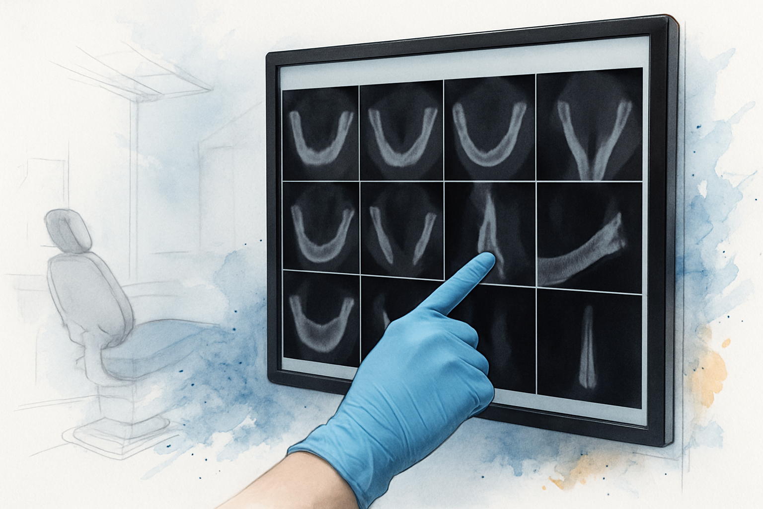
Flat X-Ray vs. 3D Scan: What Each One Can Actually Tell You
Both images have a job. This is where each one stops being useful for implant planning.
| What needs to be known | Standard flat X-ray | 3D cone beam scan |
|---|---|---|
| Bone height at the site | Approximate, can be distorted by angle | Measured directly in millimeters |
| Bone width of the ridge | Not visible at all | Visible in cross-section |
| Lower jaw nerve path | General area only | Traced along its full route |
| Sinus floor position (upper jaw) | Rough outline | Exact distance to the ridge |
| Angle available for placement | Cannot be determined | Simulated before anything is placed |
| Decay and existing restorations | Very good | Also visible, less detail on fine decay |
The Three Answers a Scan Can Produce
Once the measurements are in, the recommendation lands in one of three places. There is no fourth option.
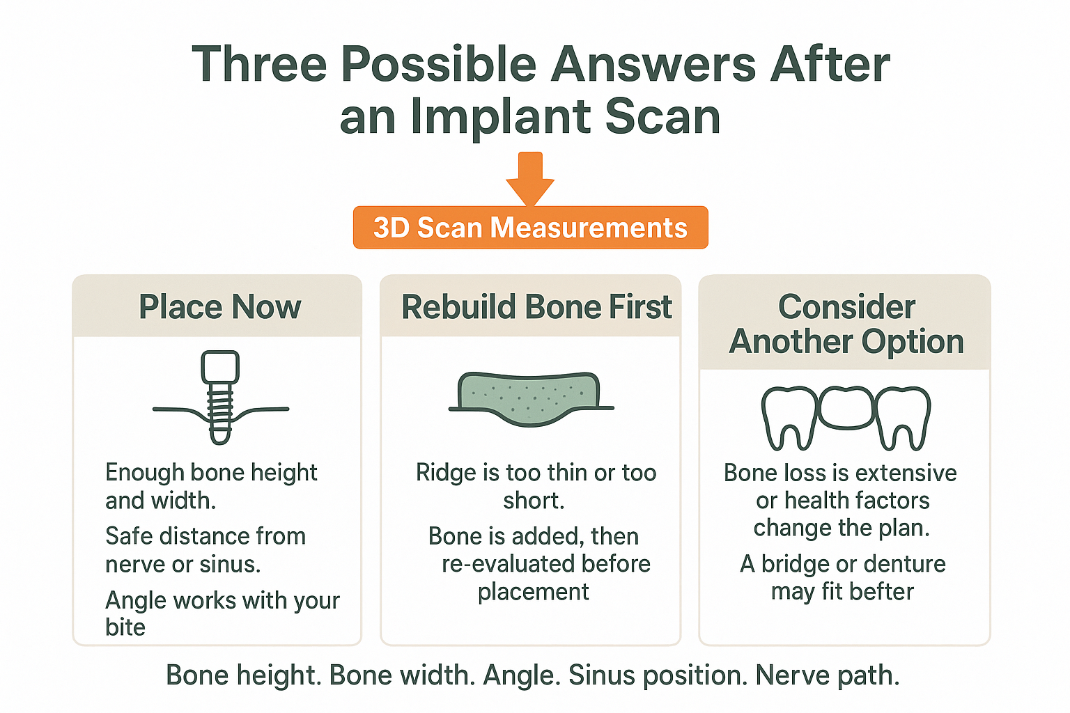
How Those Measurements Turn Into a Recommendation
Place the implant now. The bone has enough height and width, the nerve or sinus sits at a safe distance, and the angle lines up with your bite. This is the shortest path, and it is more common when a tooth was lost recently.
Rebuild the bone first. The ridge is too thin, too short, or both. Bone is added at that site, it is given time to mature, and then the site is re-measured before anything is placed.
Consider a different way to replace the tooth. Sometimes the bone loss is extensive enough, or the site complicated enough, that a bridge or a partial denture is the more sensible plan. That is not a failure. It is a recommendation based on what is actually there. If you are weighing those paths, Crown vs. Bridge vs. Implant walks through how patients tend to make that call.
One more thing worth knowing: bone starts changing shape soon after a tooth comes out, and it keeps changing quietly for years. That is why the answer for someone who lost a molar eight months ago often looks different from the answer for someone who lost one in 2015. We covered that pattern in how long you can wait after losing a tooth.
Socket Preservation, Ridge Augmentation, Gum Grafting: Four Different Procedures
Patients hear these words used interchangeably all the time, and they are not the same thing. If you can keep them straight, you can follow your own treatment plan without guessing.
- Socket preservation. Done at the time of an extraction. Grafting material goes into the empty socket right away to limit how much the bone collapses afterward. It is preventive, and it makes future implant planning simpler.
- Ridge augmentation. Done at a site where bone has already been lost. It restores width or height so there is enough structure to hold an implant. This is the one people usually mean when they say “bone graft.”
- Gum grafting. This has nothing to do with bone volume. It addresses receded gum tissue and exposed tooth roots, using healthy tissue to rebuild the gumline and reduce sensitivity.
- Periodontal surgery. This treats advanced gum disease, including deep pockets and infection under the gumline that a deep cleaning cannot reach.
A treatment plan can include more than one of these. But they solve different problems, and the scan is what tells us which problem you actually have.
If grafting turns out to be part of your plan, the sequencing question comes next, and we handle that separately in how long the implant process actually takes.
What to Ask When Someone Reviews the Scan With You
A lot of people who reach this topic have been away from dental care for a while, or had an experience elsewhere that left them wary. What settles most of them down is not reassurance. It is being shown the images and told what the clinician is looking at.
One of our patients put it plainly in a review: “She shows me my xrays, tells me what she’s looking at, and compares previous xrays, and takes lots of photos.” That is the standard to hold any office to, including ours.
When your scan comes up on the screen, ask these:
- What does the bone look like at this specific site, in height and in width?
- How close is the nerve or the sinus, and how does that affect placement?
- What would change this recommendation, either for better or worse?
- What happens if I wait six months, or a year?
- If bone needs rebuilding, is it socket preservation or ridge augmentation, and why?
If you cannot get a plain-language answer to those, that is useful information too. Anyone who is nervous about the visit itself may also find how to tell if a practice is built for anxious patients worth reading first.
Frequently Asked Questions About Implant Candidacy Scans
Do I need a 3D scan just to find out if I am a candidate?
For implant planning, yes, in almost every case. A flat X-ray cannot show ridge width or trace the nerve path, and those are two of the measurements the decision rests on. Skipping it means planning around a guess.
Can I bring a scan from another office?
Often, yes. Bring the actual image file if you can, not just a printout or a report. Depending on how old it is and how much of the jaw it captured, it may still be usable, or it may need to be repeated.
If the scan says I need bone rebuilt, does that mean I am not a candidate?
Usually it means not yet, rather than no. Ridge augmentation exists precisely for sites that have lost width or height. The scan is what determines how much needs to be rebuilt and whether that is realistic at your site.
Will my insurance cover the scan?
It varies quite a bit by plan. Some PPO plans in Washington apply diagnostic imaging benefits to it, others treat it separately, and coverage can also depend on how the rest of your treatment is coded. Our team can check your specific benefits before you commit to anything, and how dental insurance works in Washington covers the broader gaps.
How long does the scan itself take?
The imaging portion takes well under a minute. You stand or sit still while the arm rotates around your head. Nothing goes in your mouth and nothing touches you. Reviewing the results together takes far longer than capturing them.
Want to Know What Your Own Scan Would Show?
Patients driving in from Kent, Tukwila, Burien and Newcastle often assume surgical planning means a trip into Seattle. Here, the bone evaluation and any rebuilding work are handled in-house by Dr. Jaewon Kim, a board-certified periodontist, so the person reading your scan is the same person who would perform the surgical portion. That continuity is the main reason the plan tends to hold together from the first appointment forward. If you would like your own situation looked at, Cedar Dental Group is a few minutes from downtown Renton at 425-430-0400, or you can read more at cedardentalgroup.com.
